Recent Blogs

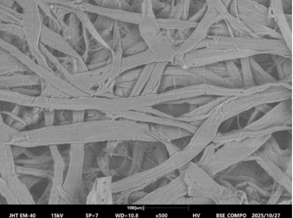
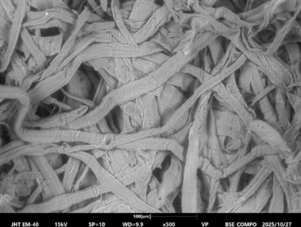
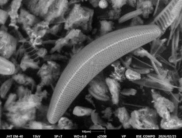
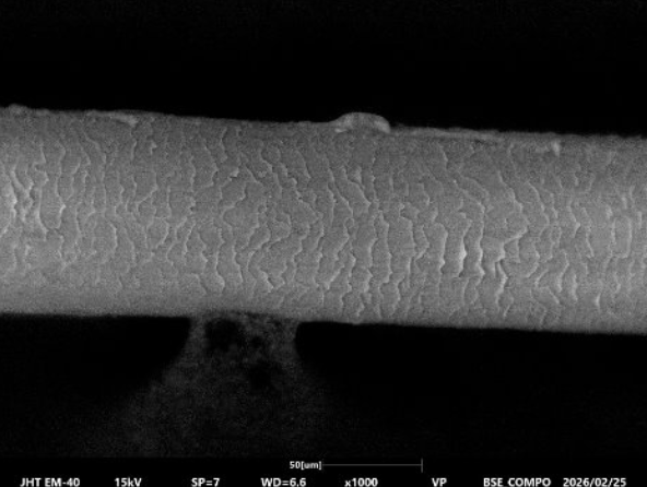
SEM: Low Vaccuum vs. High Vacuum Imaging – Webinar
Recent Blogs 5 Key Takeaways from Our Webinar: Low Vacuum

What Happens When Elves Discover Surface Metrology?
Recent Blogs What Happens When Elves Discover Surface Metrology? Candy

2025 Equipment Tax Benefits to Consider
Recent Blogs Equipment Tax Benefits for 2025! Is Now the

Reviving Legacy AFMs: How to Unlock New Capabilities
Recent Blogs Reviving Legacy AFMs: How to Unlock New Capabilities

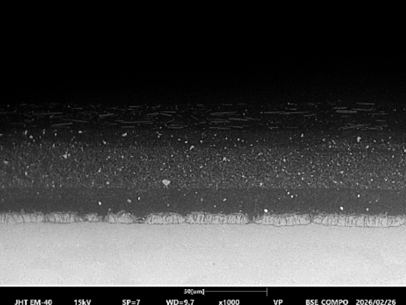
Automotive Paint Layer Analysis with Microscopy & EDS | JH Technologies
Recent Blogs Automotive Paint Layer Analysis with Optical Microscopy, SEM,
5 Key Takeaways from Our Webinar: Low Vacuum vs. High Vacuum SEM Imaging
On March 4, JH Technologies hosted a live webinar exploring one of the most common questions in scanning electron microscopy: when should you use Low Vacuum vs. High Vacuum SEM imaging?
Led by SEM experts Yi Zhang, PhD, and Ken Hirscht, the session walked attendees through practical imaging scenarios, sample preparation considerations, and a live demonstration using the COXEM EM‑40 Tabletop SEM.
If you missed the live event, the full recording is free to watch below, along with the detailed slide deck containing additional information and example images.
Why This Topic Matters
Scanning electron microscopy delivers high-resolution imaging and detailed surface analysis, but sample type and preparation can dramatically affect image quality.
While high vacuum SEM provides the highest possible resolution, it often requires conductive samples or additional preparation steps such as sputter coating.
Low vacuum imaging, on the other hand, allows researchers to analyze non-conductive samples with minimal preparation, reducing time and potential sample damage.
This webinar explored how to balance resolution, preparation time, and sample integrity when choosing between the two modes.
Key Takeaways from the Webinar
1. High Vacuum SEM Delivers Maximum Resolution
High vacuum conditions provide the most stable imaging environment and enable nanometer-scale resolution.
However, non-conductive samples can create charging effects, which distort images and obscure surface details.
“The most common SEM imaging condition is high vacuum, and it provides maximum resolution.”
— Ken Hirscht
2. Low Vacuum Imaging Reduces Charging on Non-Conductive Samples
Low vacuum SEM introduces a small amount of gas into the chamber, allowing electrons to dissipate and reducing charging artifacts.
This enables imaging of non-conductive samples without sputter coating.
“Low vacuum imaging provides extra pathways for electrons to dissipate, which helps eliminate charging effects.”
— Ken Hirscht
3. Low Vacuum Can Eliminate Sample Preparation Steps
Traditional SEM preparation often requires sputter coating or other treatments.
Low vacuum imaging can allow researchers to skip coating entirely, saving time and avoiding the need for additional equipment.
Example materials discussed in the webinar included:
Paper fibers
Human hair
Diatom algae
Automotive paint cross-sections
4. Low Vacuum Preserves Delicate Surface Features
For porous or delicate materials, coating can sometimes alter or obscure surface morphology.
Low vacuum imaging helps preserve the original surface structure.
“Since we didn’t coat the sample, the low vacuum SEM preserves the original surface features.”
— Yi Zhang
5. Faster Turnaround for Real-World Analysis
By removing coating steps and reducing preparation requirements, low vacuum imaging can significantly speed up analytical workflows.
This is especially useful for:
Failure analysis
Rapid materials characterization
Industrial quality control
“Low vacuum imaging allows faster turnaround for quick analysis.”
— Yi Zhang
Live Demo Highlights
During the session, Yi and Ken demonstrated imaging of an automotive paint cross-section sample using the COXEM EM-40 tabletop SEM.
The demo compared:
High vacuum (coated sample)
High vacuum (uncoated sample)
Low vacuum (uncoated sample)
Attendees were able to see how low vacuum imaging successfully revealed multiple paint layers and embedded particles without coating.
Key features shown during the demo included:
Adjustable low vacuum pressures (10–30 Pa)
BSE detector imaging
Rapid autofocus and image capture
Software navigation and stage control
Â
Interested in learning more about SEM imaging strategies?
âś” Watch the full webinar recording below
âś” Download the detailed slide deck below
âś” Contact JH Technologies to discuss SEM solutions for your lab
Learn more about SEM systems available through JH Technologies or explore the capabilities of the COXEM EM‑40 Tabletop SEM.Â
Connect with our team and request a quote today!
Webinar Video